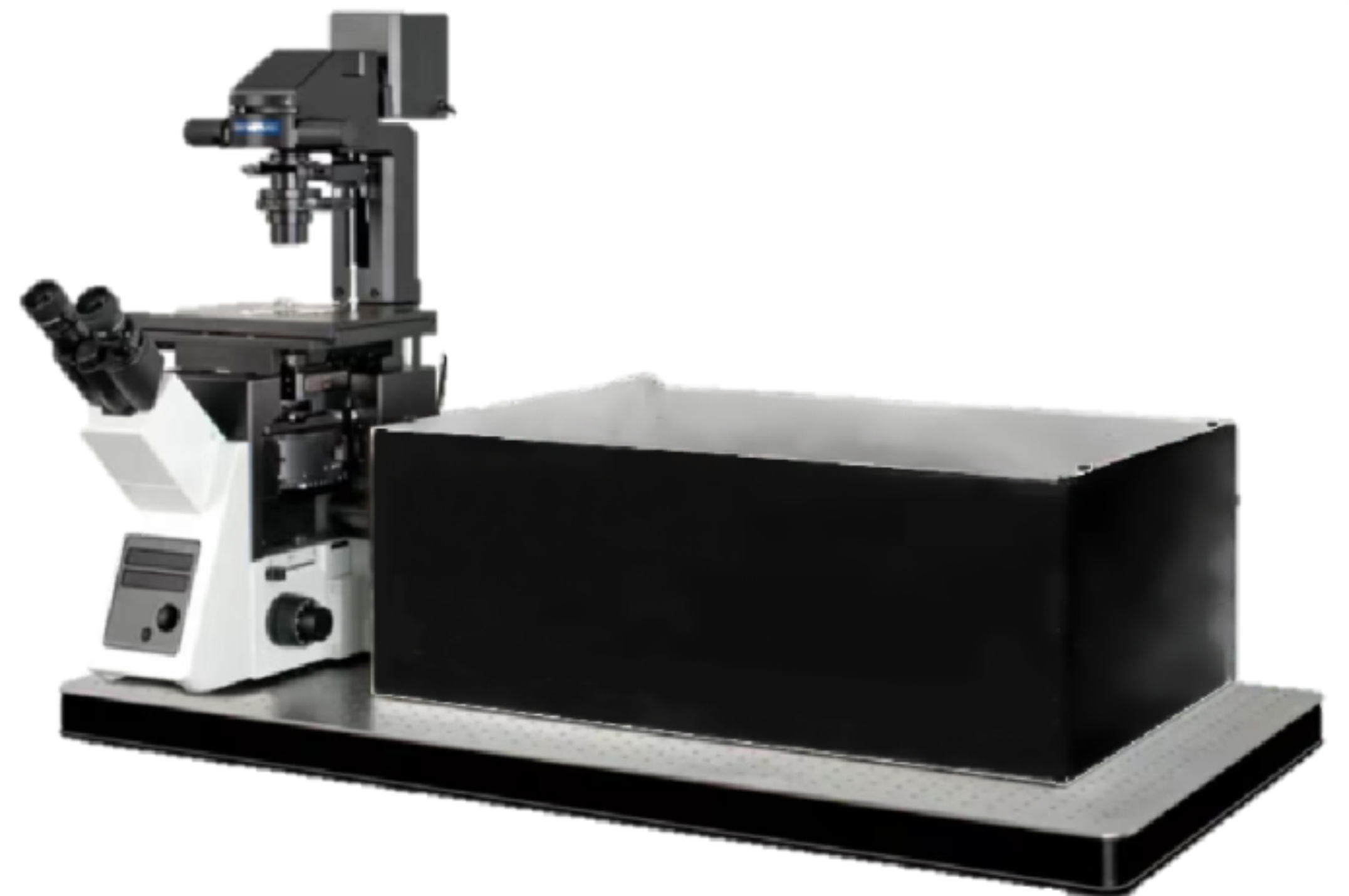
亚细胞光片扫描显微镜
产品名称: 亚细胞光片扫描显微镜
英文名称: 亚细胞光片扫描显微镜
产品编号: LSI LBS系列
产品价格: 0
产品产地: 中国
品牌商标: LogiSci
更新时间: 2024-12-12T13:37:58
使用范围: null
- 联系人 :
- 地址 : 武汉东湖高新区光谷大道特1号国际企业中心三期1栋
- 邮编 : 430070
- 所在区域 : 湖北
- 电话 : 133****6638 点击查看
- 传真 : 点击查看
- 邮箱 : aphotonic@163.com
<p><p style="margin-left:0pt; margin-right:0pt; text-align:left"><span style="font-size:16px"><span style="color:#231f20">LSI系列激光片层扫描显微镜以前所未有的灵敏度,分辨 率以及成像速度帮助生物学家解读活体样品的三维动态 过程。LSI系列显微镜使用了最前沿的光学和工程技术来 产生一束超薄的线性贝塞尔片层光,并用它来实现对生 物样品的高精度光学层析。此项专利技术的应用不仅显 着提高了片层光显微系统的成像分辨率,而且允许系统 使用超弱的激发光便可从样品中获得足够的信号强度, 所以极大的减弱了样品在成像时承受的光毒性,延长了 样品的有效观测时间,以此帮助观测者获得更多高质量的成像数据。 </span></span></p> <p style="margin-left:0pt; margin-right:0pt; text-align:left"> </p> <p style="margin-left:0pt; margin-right:0pt; text-align:center"><span style="font-size:16px"><img alt="" src="https://nwzimg.wezhan.cn/contents/sitefiles2053/10267688https://msimg.bioon.com/bionline//images/28597745.jpg" style="height:415px; width:554px"></span></p> <p style="margin-left:0pt; margin-right:0pt; text-align:center"> </p> <p style="margin-left:0pt; margin-right:0pt; text-align:justify"><span style="font-size:16px"><strong><strong>超越激光共聚焦显微技术</strong></strong></span></p> <p style="margin-left:0pt; margin-right:0pt; text-align:justify"><span style="font-size:16px">LSI系列显微镜将激发光的能量严格限制在中心厚度不到400纳米的片层光中。片层光与探测物镜的焦平面重合,用来激发仅在探测景深范围内的样品结构,因此在成像时不会产生任何的背景噪声。</span></p> <p style="margin-left:0pt; margin-right:0pt; text-align:justify"><span style="font-size:16px">同时配合探测物镜具有超大数值孔,可以高效的接收样品发出的微弱荧光信号,且产生的图像可达光学极限分辨率。相较与共聚焦显微,LSI系列在以下方面具有显着优势:</span></p> <p style="margin-left:0pt; margin-right:0pt; text-align:justify"> </p> <p style="margin-left:0pt; margin-right:0pt; text-align:justify"><span style="font-size:16px"><strong><strong>高速活细胞成像</strong></strong> <strong><strong>超低的光毒性</strong></strong></span></p> <p style="margin-left:0pt; margin-right:0pt; text-align:justify"><span style="font-size:16px">★拍摄速度可达500幅每秒 ★相较共聚焦减弱1000倍!</span></p> <p style="margin-left:0pt; margin-right:0pt; text-align:justify"> </p> <p style="margin-left:0pt; margin-right:0pt; text-align:justify"><span style="font-size:16px"><strong><strong>高分辨率三维结构成像 </strong></strong> LBS激光片层扫描显微系统</span></p> <p style="margin-left:0pt; margin-right:0pt; text-align:justify"><span style="font-size:16px">★250nm横向分辨率 开创了五维活细胞生物成像的时代:</span></p> <p style="margin-left:0pt; margin-right:0pt; text-align:justify"><span style="font-size:16px">★350nm轴向分辨率 <strong><strong>★</strong></strong><strong><strong>3维空间+1维时间+1维颜色</strong></strong></span></p> <p style="margin-left:0pt; margin-right:0pt; text-align:justify"> </p> <p style="margin-left:0pt; margin-right:0pt; text-align:center"><span style="font-size:16px"><strong><strong><img alt="" src="https://nwzimg.wezhan.cn/contents/sitefiles2053/10267688https://msimg.bioon.com/bionline//images/28597746.jpg" style="height:505px; width:554px"></strong></strong></span></p> <p style="margin-left:0pt; margin-right:0pt; text-align:center"><span style="font-size:16px">LSI系列片层扫描显微系统的成像原理示意简图</span></p> <p style="margin-left:0pt; margin-right:0pt; text-align:center"> </p> <p style="margin-left:0pt; margin-right:0pt; text-align:justify"><span style="font-size:16px"><strong><strong>超越传统激光片层扫描显微技术</strong></strong></span></p> <p style="margin-left:0pt; margin-right:0pt; text-align:justify"><span style="font-size:16px"><span style="color:#231f20">传统的片层光显微技术普遍通过扫描汇聚的高斯光束或者使用柱面镜压缩一个准直的高斯头束来产生片层光而这两种方式产生的片层光在厚度和长度皆被光的衍射特性限制。而LBS技术通过一系列光学手段则可以打破这一限制:产生更薄且更长的LSI系列片层光。因此LSI系列系统在保持传统片层扫描显微技术具有的高成像速度和低光毒性优势的同时,凭藉更精细的光学层析能力进一步显着地提高了成像分辨率和灵敏度。</span></span></p> <p style="margin-left:0pt; margin-right:0pt"> </p> <p style="margin-left:0pt; margin-right:0pt; text-align:center"><span style="font-size:16px"><img alt="" src="https://nwzimg.wezhan.cn/contents/sitefiles2053/10267688https://msimg.bioon.com/bionline//images/28597748.jpg" style="height:110px; width:554px"></span></p> <p style="margin-left:0pt; margin-right:0pt; text-align:center"> </p> <p style="margin-left:0pt; margin-right:0pt; text-align:left"><span style="font-size:16px">通过扫描或者用柱面镜压缩一个高斯光束得到的薄(但长度不足)或者长(但过厚)的片层光</span></p> <p style="margin-left:0pt; margin-right:0pt; text-align:justify"><span style="font-size:16px">LSI系列系统产生的超薄且长的LSI系列片层光</span></p> <p style="margin-left:0pt; margin-right:0pt; text-align:justify"> </p> <p style="margin-left:0pt; margin-right:0pt; text-align:center"><span style="font-size:16px"><img alt="" src="https://nwzimg.wezhan.cn/contents/sitefiles2053/10267688https://msimg.bioon.com/bionline//images/28597749.jpg" style="height:271px; width:554px"></span></p> <p style="margin-left:0pt; margin-right:0pt; text-align:center"> </p> <p style="margin-left:0pt; margin-right:0pt"><span style="font-size:16px">相较于传统片层光显微系统,LSI系列技术显着提高了成像系统的光学层析能力和图片的信噪比。比例尺:3微米</span></p> <p style="margin-left:0pt; margin-right:0pt"> </p> <p style="margin-left:0pt; margin-right:0pt"><span style="font-size:16px"><strong><span style="color:#231f20"><strong>亚细胞分辨多维光片成像系统</strong></span></strong></span></p> <p style="margin-left:0pt; margin-right:0pt; text-align:justify"> </p> <p style="margin-left:0pt; margin-right:0pt; text-align:justify"><span style="font-size:16px"><strong><strong>高度集成的设计</strong></strong></span></p> <p style="margin-left:0pt; margin-right:0pt; text-align:justify"><span style="font-size:16px">LSI系列片层扫描显微镜可立即用于活细胞成像实验:每台显微镜都集成的一套活细胞培养(灌注)系统,这一系统配有精确的温度/二氧化碳环境控制模块从而实现长时间活细胞成像;同时集成了一套具有大视野的EPI荧光显微模块用于定位拍摄目标;以及一套可达纳米精度的三维电动样品台,和最多可集成6通道的Solar2.0光纤激光模块作为光源</span></p> <p style="margin-left:0pt; margin-right:0pt; text-align:justify"> </p> <p style="margin-left:0pt; margin-right:0pt; text-align:justify"><span style="font-size:16px"><strong><strong>具有温度/C02控制的活细胞样品灌注池</strong></strong></span></p> <p style="margin-left:0pt; margin-right:0pt; text-align:justify"><span style="font-size:16px">◆可注入2-5ml培养液或任何液体用于浸润样品</span></p> <p style="margin-left:0pt; margin-right:0pt; text-align:justify"><span style="font-size:16px">◆可实现拍摄时更换培养液或加入药物</span></p> <p style="margin-left:0pt; margin-right:0pt; text-align:justify"><span style="font-size:16px">◆集成了一个Epi荧光成像通道,可选配4x/10x/50x空气物镜</span></p> <p style="margin-left:0pt; margin-right:0pt; text-align:justify"> </p> <p style="margin-left:0pt; margin-right:0pt; text-align:center"><span style="font-size:16px"><img alt="" src="https://nwzimg.wezhan.cn/contents/sitefiles2053/10267688https://msimg.bioon.com/bionline//images/28597750.jpg" style="height:580px; width:554px"></span></p> <p style="margin-left:0pt; margin-right:0pt; text-align:center"> </p> <p style="margin-left:0pt; margin-right:0pt; text-align:justify"><span style="font-size:16px"><strong><strong>最大化的适用范围</strong></strong></span></p> <p style="margin-left:0pt; margin-right:0pt; text-align:justify"><span style="font-size:16px">LSI系列激光片层扫描显微镜可适用于不同种类与大小的样品。可观测的样品范围包括了细胞爬片,酵母菌细胞或植物细胞组织等。加装大样品成像模块后可将应用扩展至胚胎、小型动物如线虫,果蝇幼虫或者斑马鱼的观测</span></p> <p style="margin-left:0pt; margin-right:0pt; text-align:justify"> </p> <p style="margin-left:0pt; margin-right:0pt; text-align:justify"><span style="font-size:16px"><strong><strong>应用实例</strong></strong></span></p> <p style="margin-left:0pt; margin-right:0pt; text-align:justify"> </p> <p style="margin-left:0pt; margin-right:0pt; text-align:center"><span style="font-size:16px"><strong><strong><img alt="" src="https://nwzimg.wezhan.cn/contents/sitefiles2053/10267688https://msimg.bioon.com/bionline//images/28597751.jpg" style="height:202px; width:554px"></strong></strong></span></p> <p style="margin-left:0pt; margin-right:0pt; text-align:center"><span style="font-size:16px"><span style="color:#231f20">LSI系列片层扫描显微系统拍摄的细胞中微管(绿色)和线粒体(红色)结构的三维荧光显微图像</span></span></p> <p style="margin-left:0pt; margin-right:0pt"> </p> <p style="margin-left:0pt; margin-right:0pt; text-align:center"><a href="https://msimg.bioon.com/bionline/ewebeditor/uploadfile/202207/20220729114724316.png" target="_blank"><img src="https://msimg.bioon.com/bionline/ewebeditor/uploadfile/202207/20220729114724316_s.png" border="0"></a><br></p> <p><br></p></p>
